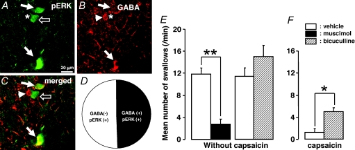
Figure 8. GABA-LI neurons or pERK-LI neurons in the NTS of the rats with 10 mm capsaicin injection to the lingual muscle and the effect of muscimol or bicuculline microinjection into the NTS on swallowing reflex.
A, pERK-LI neurons in the NTS; B, GABA-LI neurons in the NTS; C, pERK-LI neurons with GABA immunoreactivity; D, pie chart showing the distribution of the GABA-LI and GABA-negative pERK-LI neurons in the NTS. White arrows in A indicate pERK-LI neurons, those in B indicate GABA and those in C indicate GABA and pERK-LI neurons. The open arrows in A and C indicate pERK-LI neuron without GABA immunoreactivity. The arrowhead in B indicates pERK negative/GABA positive. The star in A corresponds to the arrowhead in B and C, and that in B corresponds open arrows in A and C. E and F, mean number of swallows in vehicle-treated rats and that following microinjection of muscimol or bicuculline to the NTS, and that following microinjection of vehicle or bicuculline to the NTS in capsaicin-treated rats. *P < 0.05, **P < 0.01.

