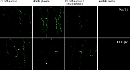
Figure 2. Immunocytochemistry of PepT1 and PLC β2 regulation by glucose and artificial sweeteners in rat jejunum.
Rat jejunum was perfused with 75 mm glucose, 20 mm glucose or 20 mm glucose and 1 mm sucralose for 30 min. Sections (7 μm) were labelled with a rabbit primary antibody detecting either PepT1 or PLC β2 followed by a goat anti-rabbit secondary antibody conjugated to Alexa 488 (green). Arrows: apical membrane; arrowheads: three different types of SCC containing PLC β2. The peptide controls were obtained by neutralization of primary antibody with excess immunogenic peptide. Images were processed using spectral unmixing techniques (see Methods), which automatically subtract all background contributions to leave only specific labelling as demonstrated by the peptide controls, which are totally black.

