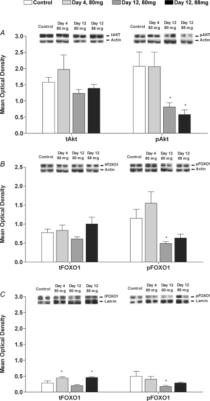
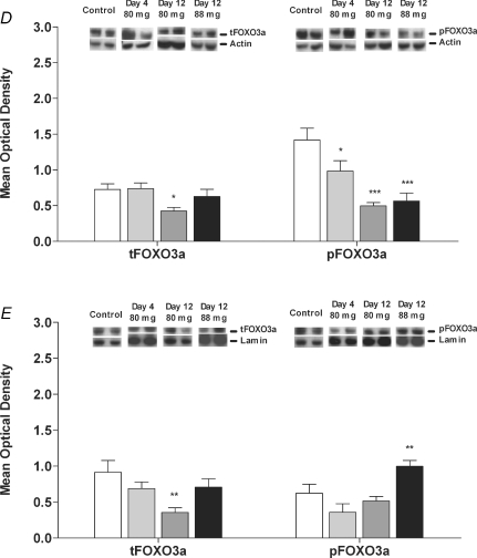
Figure 1. Protein expression of Akt, FOXO1 and FOXO3a in biceps femoris muscle of Control animals and animals dosed daily with 80 mg kg−1 day−1 simvastatin for 4 and 12 days and 88 mg kg−1 day−1 simvastatin for 12 days.
A, total Akt (tAkt) and phosphorylated Akt (pAkt) in the cytosolic protein fraction; B, total FOXO1 (tFOXO1) and phosphorylated FOXO1 (pFOXO1) in the cytosolic protein fraction; C, total FOXO1 (tFOXO1) and phosphorylated FOXO1 (pFOXO1) in the nuclear protein fraction; D, total FOXO3a (tFOXO3a) and phosphorylated FOXO3a (pFOXO3a) in the cytosolic protein fraction; E, total FOXO3a (tFOXO3a) and phosphorylated FOXO3a (pFOXO3a) in the nuclear protein fraction. Phosphorylated to total protein ratios are described in Results. Values expressed as mean optical density ±s.e.m.*P < 0.05, **P < 0.01, ***P < 0.001 when compared to Control. Images show a representative Western blot of the protein of interest and the corresponding actin or lamin blot.


