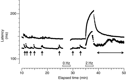
Figure 1. Microneurographic raster plot of latencies in the C fibre range during baseline stimulation (0.25 Hz), pause (0 Hz, first open bar) and tetanus (2Hz, second open bar) from a patient with small fibre neuropathy and spontaneous burning pain.
Two different fibres with activity-dependent profiles characteristic of mechano-insensitive nociceptors can be observed (baseline latency 123 ms and 150 ms, respectively). Both units were spontaneously active, giving rise to some ‘saw-tooth’ markings (marked with arrows for the fibre at a shorter latency) before the pause. After the 2 Hz tetanus the fibre at a shorter latency engages in intense spontaneous activity with progressive increase of conduction latency (horizontal double-ended arrow).

