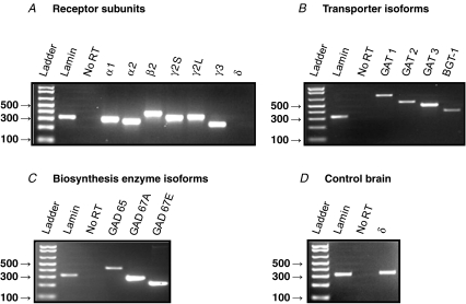
Figure 7. RT-PCR analysis of GABAergic marker expression in extracts of postnatal day (P) 14 rat petrosal ganglia.
Products were run on 2% agarose gel stained with ethidium bromide and viewed under UV illumination. A, expression of GABAA receptor subunits; note lack of expression of the δ subunit. B, expression of the indicated GABA transporter (GAT) isoforms, including BGT-1. C, expression of the indicated GABA biosynthetic enzyme, glutamic acid decarboxylase (GAD) isoforms. D, positive expression of GABAAR δ subunit in rat brain tissue verifies primer function. Lamin (A/C) housekeeping gene was used as a positive control. ‘No RT’ lane refers to negative control reactions in which water replaced Superscript III reverse transcriptase. All band identities were verified by sequencing (MOBIX, McMaster University, Canada).

