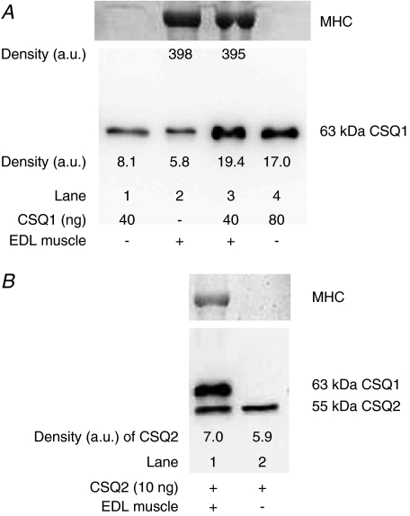
Figure 2. Verification of CSQ quantification by adding pure CSQ to muscle homogenates.
A, lower panel shows Western blot (probed with anti-CSQ1) of lanes containing purified CSQ1 alone (40 ng, lane 1; 80 ng, lane 4), EDL muscle homogenate alone (lane 2), or the same amount of muscle homogenate with 40 ng added CSQ1 (lane 3). Post-transfer Coomassie Blue staining of the gel for MHC (upper panel) verified that lanes with muscle homogenate had a similar amount loaded. Density values for CSQ1 and MHC are indicated. Band densities indicate that the mixture of purified CSQ1 and muscle homogenate CSQ1 was transferred, detected and quantified with similar efficacy as purified CSQ1 run by itself (also see Results). B, similar to A, but with 10 ng pure CSQ2 added to lanes 1 and 2 and EDL muscle homogenate present in lane 1 only (probed with anti-CSQ1 and 2). The EDL muscle contains virtually no CSQ2, and it is apparent that the detection of the exogenous CSQ2 was not hindered by the proteins present in the whole muscle sample; CSQ1 was also detected in the muscle sample.

