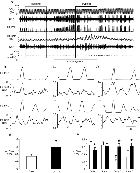
Figure 1. Effects of hypoxia on phrenic and splanchnic sympathetic nerve activity.
A, raw tracings of end-tidal CO2, phrenic nerve discharge (PND), splanchnic sympathetic nerve activity (SNA), integrated (Int.) nerve activities, and arterial pressure (AP) at baseline and during the hypoxia protocol. Ba, Ca and Da, phrenic-triggered histograms from 3 rats to display SNA in relation to central respiratory drive (made with 30 s of baseline SNA and PND). Bb, Cb and Db, the final 30 s of activity during 60 s of 10% O2 inhalation was used to evaluate SNA in relation to PND during hypoxia in the same 3 rats from Ba, Ca and Da. E, mean SNA collected for 30 s at baseline and during hypoxia (inside boxes in A). F, mean SNA was evaluated during the early inspiratory (Early I), late inspiratory (Late I), early expiratory (Early E) and late expiratory (Late E) phases of respiration. Open bars represent SNA at baseline. Filled bars represent SNA during hypoxia. *Significant difference from baseline value during that period. †Significant difference from values during both phases of inspiration.

