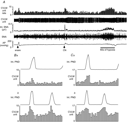
Figure 2. Examples of baro-activated caudal ventrolateral medulla (CVLM) neurones activated by hypoxia.
A, this CVLM neurone was activated by raising AP (snare), i.v. injection of sodium cyanide (CN), and inhalation of hypoxic air (bar = 60 s of hypoxia). B, phrenic-triggered histogram of CVLM neuronal activity using 30 s of baseline activity (Ba) and 30 s during hypoxia (Bb). C, phrenic-triggered histogram of another baro-activated, hypoxia-activated CVLM neurone at baseline (Ca) and during hypoxia (Cb). The CVLM units were counted in 0.1 s bins.

