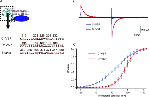
Figure 1. Topology of voltage-sensing phosphatase (VSP) and charge movements of the voltage sensor.
A, the S4 segment in the voltage sensor (dotted box) contains multiple positively charged residues (red). Amino acids from ascidian and teleost VSP (Ci-VSP and Dr-VSP) are compared with those of the Drosophila Shaker potassium channel. B, ‘gating’ or sensing currents evoked at 160 mV under whole-cell patch clamp from tsA201 cells transfected with Ci-VSP (blue) and Dr-VSP (red). C, charge–voltage (Q–V) relations of Ci-VSP (blue) and Dr-VSP (red) (modified from Hossain et al. 2008). Normalized values of Off-charges are shown as mean and standard deviations, collected from 6 and 5 cells for Ci-VSP and Dr-VSP, respectively. Maximum Off-charges were 2.89 ± 0.3 pC pF−1 and 0.11 ± 0.1 pC pF−1 for Ci-VSP and Dr-VSP, respectively.

