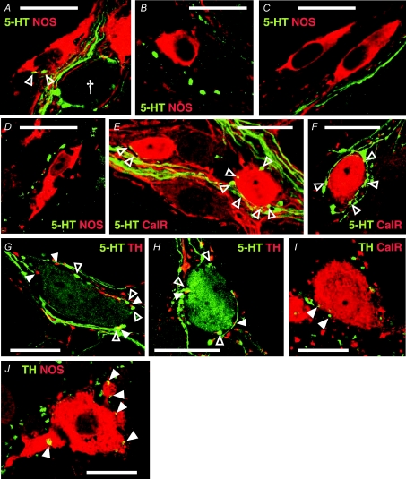
Figure 6. 5-HT and TH connections to jejunal neurones.
A–D. 5-HT-IR (green) varicosities apposed to NOS-IR (red) cell bodies (open arrowheads) were extremely rare in both Balb/c (A and B) and C57Bl/6 (C and D) mice. Occasionally rings of 5-HT-IR varicosities were observed surrounding NOS−ve cell bodies (A, †), but never surrounding NOS-IR cell bodies. Many NOS-IR neurones were not apposed by any 5-HT-IR varicosities (B–D). E and F, 5-HT-IR varicosites in apposition to calretinin-IR (red) neurones were relatively abundant in jejunum samples from both strains. Calices of varicosities arising from passing 5-HT-IR axons (E) were often seen in apposition to calretinin-IR cell bodies. Rings (F) of 5-HT-IR varicosities were also frequently observed surrounding calretinin-IR cell bodies. G and H, 5-HT-IR cell bodies were apposed by numerous 5-HT-IR and TH-IR (red, filled arrowheads) varicosities in Balb/c (G) and C57Bl/6 (H) jejunum. I and J, TH-IR varicosities (green) were observed in apposition to both calretinin-IR (red, I) and NOS-IR (red, J) cell bodies (filled arrowheads) in jejunum. These images were constructed by merging 1–3 planes from z-series. Scale bars, 20 μm.

