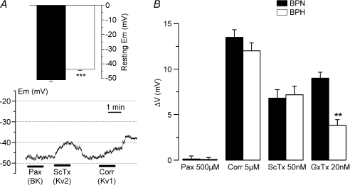
Figure 9. Resting EM in BPN and BPH VSMCs.
A, values of resting EM obtained in current-clamp experiments with the perforated-patch configuration from BPH and BPN mesenteric VSMCs; n= 39. The lower graph shows resting EM values in one BPN cell during the application of 500 nm paxilline, 50 nm ScTx and 5 μm correolide as indicated with the bars. B, the average depolarization (means ±s.e.m.) obtained in 9–15 BPN and BPH cells with the different drugs explored is represented as ΔVm.

