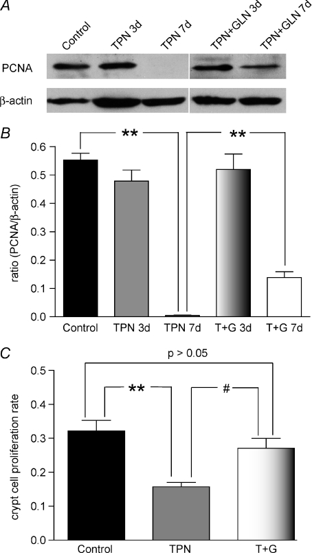
Figure 9. Proliferating cell nuclear antigen (PCNA) expression.
A, representative bands of PCNA and β-actin, which was used for normalization. B, results of Western immunoblotting as expressed as the ratio of the PCNA to β-actin. A slight decline was noted at 3 days, and a this decline became significant after 7 days of TPN administration. Glutamine supplemented TPN prevented this decline at 3 days, and partially prevented the decline at day 7. C, crypt cell proliferation is shown as detected by the counting of BrdU positive staining. TPN again led to a significant decline in proliferation compared to control mice; and glutamine supplemented TPN (T + G) prevented this decline in proliferation rates. Results for BrdU staining are shown for day 7. #P < 0.05, **P < 0.001; n= 4–6 per group.

