FIGURE 2.
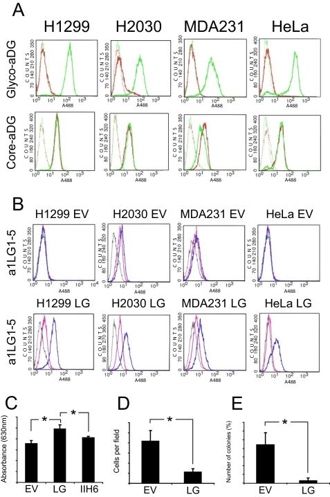
Effects of LARGE expression in cell lines expressing hypoglycosylated α-dystroglycan. A, after cancer cell lines with stable expression of LARGE were generated, the modified cells were tested by FACS using antibody IIH6 (Glyco-aDG) and antibody GT20ADG (Core-aDG). Each cell line expressing the empty virus (EV, red trace), was compared with its LARGE-expressing counterpart (LG, green trace). Solid line, primary and secondary antibody; broken line, secondary antibody only. In all cases, LARGE expression restored expression of the α-dystroglycan glyco-epitope. B, cancer cell lines that stably express LARGE were also tested by FACS analysis for the ability to bind the laminin α1-derived fusion protein a1LG1–5 in the presence of Ca2+/Mg2+ (blue trace) or EDTA (pink trace). Gray trace, secondary antibody only. LARGE expression was found to confer the ability to bind laminin α1 to the epithelium-derived cancer cells. C, assay for adhesion of cells to laminin-111. 96-well plates were coated with laminin-111, and then MDA231 EV (EV) and MDA231 LG (LG) cells were seeded at 1.5 × 10⁵ cells per laminin-coated well. Cell attachment was measured by crystal violet staining 1 h later. Attachment of MDA231 LG cells was also measured in the presence of antibody IIH6 (Glyco-aDG). The standard error (error bars) was calculated using the Student's test (n = 4); *, p < 0.01. D, Transwell migration of MDA231 cells through Matrigel-coated 8-μm pore filters. MDA-MB-231 cells were incubated in the upper chambers, in culture medium without fetal bovine serum, for 24 h. During this time, they migrated toward the bottom chamber, which contained medium with 10% fetal bovine serum. Cells were counted from at least four random fields at ×20 magnification. The standard error was calculated using the Student's test (n = 6); *, p < 0.01. E, effect of LARGE expression on anchorage-independent growth of MDA-MB-231 cells. Cells were suspended in 0.3% agar medium and layered onto a 0.5% agar base layer (n = 3). After 28 days, colony number was assessed following crystal violet staining. The standard error was calculated using the Student's test (n = 3); *, p < 0.01.

