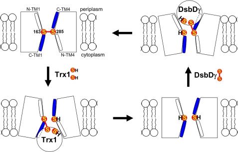
FIGURE 5.
Model of structural changes of DsbDβ during redox reactions. The model shows that DsbDβ adopts relative small openings toward both sides of the membrane when Cys-163 and Cys-285 are in the oxidized and reduced states, whereas when interacting with either partner Trx proteins, it adopts a relative large opening toward the interacting side of the membrane. S represents the sulfur of thiol in a cysteine residue. Halves of C-terminal TM1 and TM4 are water-exposed (blue; C-TM1 and C-TM4), whereas those of N-terminal ones are not (white; N-TM1 and N-TM4).

