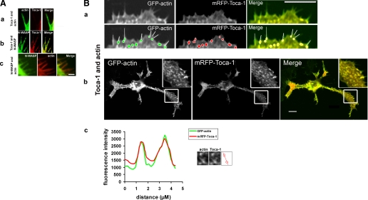
FIGURE 6.
Localization of N-WASP and Toca-1 with actin. N1E115 cells were transfected with Toca-1 with GFP-actin, Toca-1 with N-WASP, or N-WASP with mRFP-actin, and filopodia and vesicles followed. Images are in a series of three: GFP, mRFP, and overlay. A, a, localization of Toca-1/actin; b, Toca-1/N-WASP; and c, N-WASP/actin in filopodia. B, GFP-actin/mRFP-Toca-1 expression in N1E115 cells. Lower panels of a are duplicates that have tracings showing the relationship between filopodia and Toca-1 localization. Toca-1 is found at the base of filopodia. Localization of Toca-1 with actin, at the leading edge (a) and in neurites (b) (the inset is an enlargement of the box area). c, intensity profile of two of the vesicles from b showing that Toca-1 and actin co-localize. Bar = 10 μm.

