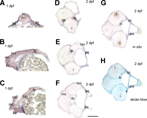
FIGURE 4.
Cryosections of 1 and 2 dpf zebrafish embryos. All sections were obtained from whole mount embryos that have undergone decorin in situ hybridization. A, cross-section through a 1 dpf embryo at the trunk level high-lighting decorin expression in the lateral plate mesoderm (lm). B and C, longitudinal sections of a 1 dpf embryo through the head region indicating decorin expression in the developing head mesenchyme (hm) and heart region. D-G, cross-sections through 2 dpf embryos at the head region, localizes decorin expression within the olfactory placode (op), trabecular cartilage (tc), placode (p), palatoquadrate (pq), quadrate cartilage (qc), and head mesenchyme (hm). H, cross-section from G was counterstained with Alcian blue correlating staining of the cartilage with regions of decorin positive expression by ISH. Spinal cord, sc; notochord, nc; mesencephalon, mc; prosencephalon, pc; placordal plate, pp; floor plate, fp; lens, l; diencephalon, dc. Bar, 250 μm.

