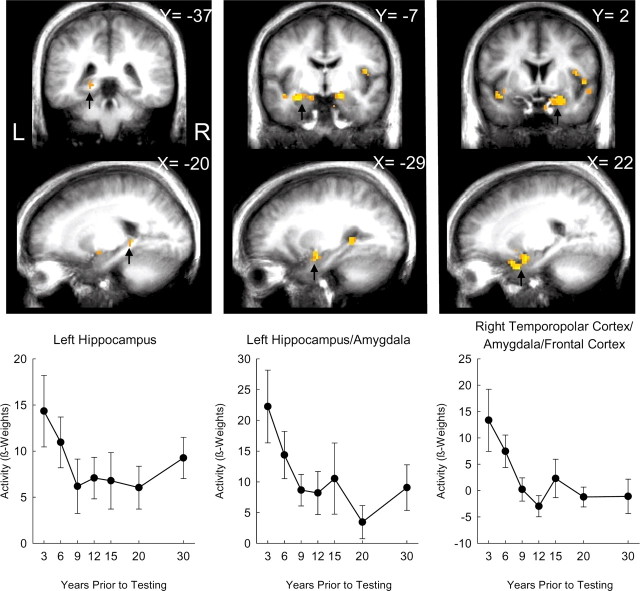
Figure 4.
Brain regions in the medial temporal lobe in which activity was related to the age of the memory. For regions identified by black arrows in the coronal sections (top row) and sagittal sections (middle row), activity levels (relative to baseline) are depicted for each time period covered by the test (bottom row). Error bars show SEM. X, Left/right Talairach coordinate; Y, anterior/posterior Talairach coordinate; L, left; R, right.

