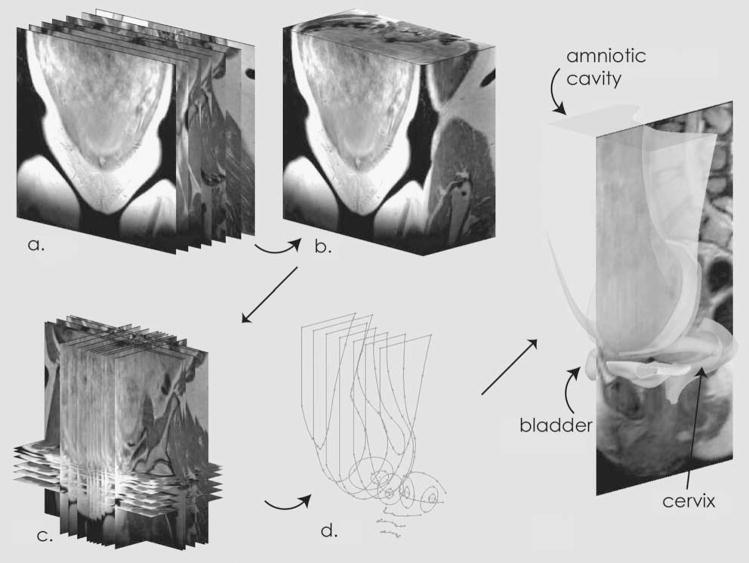
Figure 1.
Construction of Solid Models. Two-dimensional images were collected in the coronal plane (a) and converted to a three-dimensional volume (b). Individual images were selected from the three-dimensional volume and placed in the modeling workspace (c). Using these images as guides, two-dimensional tracings were made (d), which were combined (lofted) into three-dimensional solids.

