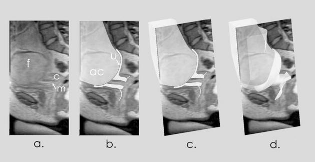
Figure 2.
Accuracy of Solid Models. The first panel (a) shows a midline, sagittal MR image with the fetal head (f), posterior cervix (c) and mucosa (m). The second panel (b) shows the anatomic model superimposed on the image. The amniotic cavity (ac) and uterus (u) are indicated. In the third panel (c), the model and image are viewed at an oblique angle. The 3D model is cut at the level of the image. The fourth panel (d) shows the full 3D anatomic model.

