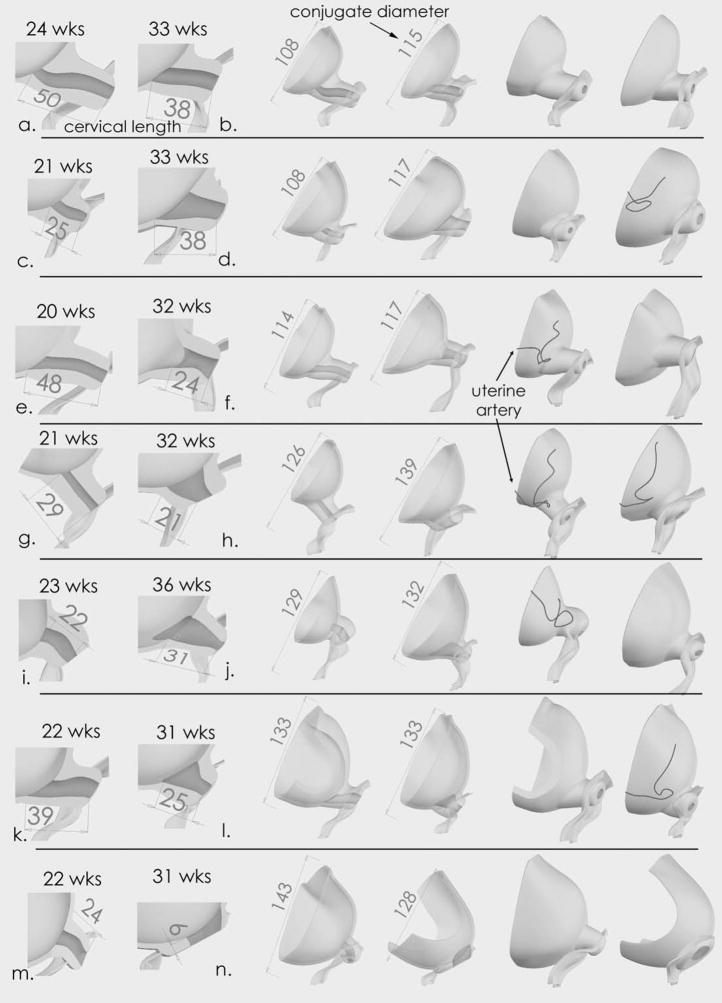
Figure 3.
Fourteen Solid Models. Each row shows two, matched models - one model from the second trimester and one from the third trimester. The models are matched by the distance of the conjugate diameter of the pelvic bone. The first two columns demonstrate cervical length. The middle two columns show an anterior view and the cervix is transparent. The last two columns show a posterior view. In all models, the volume of the amniotic cavity below the diagonal conjugate is larger in the third trimester compared to the second trimester. In addition, the transition from the cervix to the uterus is wider in the third trimester compared to the second trimester (compare (c) vs. (d), (g) vs. (h), (i) vs. (j), (k) vs. (m)). Model (n) shows a cervix that is completely effaced at 31 weeks. This patient delivered at 33 weeks. Dimensions are in millimeters.

