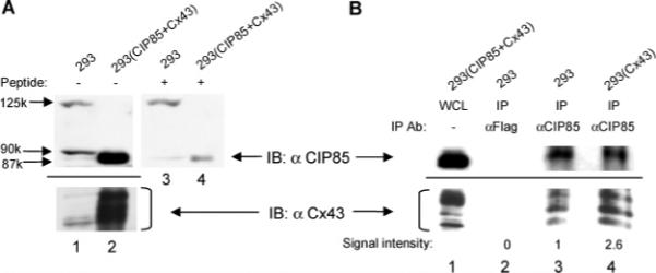
Figure 4.

Interaction between endogenous CIP85 and Cx43 in vivo. Panel A, detection of endogenous CIP85 and Cx43 in 5 mg of lysates from HEK293 cells using the CIP85 or Cx43 antibodies. The CIP85 antibody was incubated without (lanes 1 and 2) or with (lanes 3 and 4) 0.3 μg/μL CIP85 epitope peptide before immunoblotting. Lysates (0.1 mg) of HEK293 transfectants coexpressing exogenous CIP85 and Cx43 were used as positive controls (lanes 2 and 4). Panel B, nontransfected HEK293 cells or HEK293 transfectants expressing exogenous Cx43 were lysed in 300 μL of 0.2% NP-40 lysis buffer. A Flag antibody or the CIP85 antibody was used to immunoprecipitate the protein complexes, and the associated Cx43 was detected with a Cx43 antibody (lanes 2−4). Lysates of HEK293 transfectants coexpressing CIP85 and Cx43 were used as positive controls (lane 1). The increased amount of Cx43 co-immunoprecipitated with CIP85 in HEK293 cells transfected with Cx43 was quantitated by densitometry, and the signal intensity was normalized to that obtained from nontransfected HEK 293 cells for comparison.
