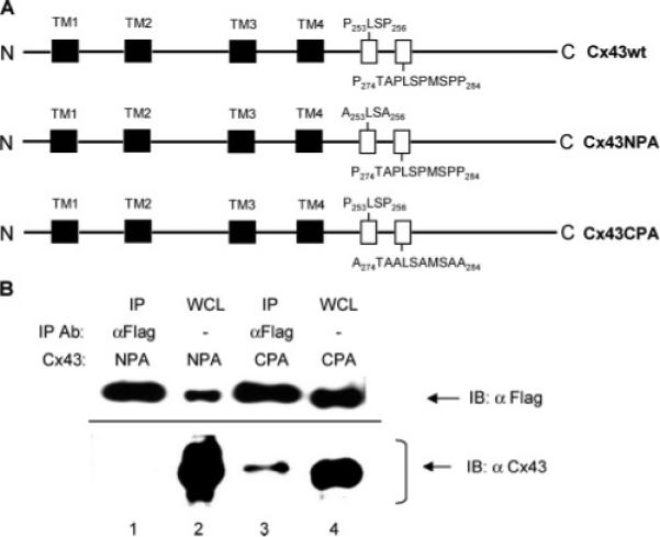
Figure 6.

Identification of the CIP85-interacting region in Cx43 by analyses of proline site substitution mutants. Panel A, schematic diagrams of the Cx43 proline to alanine substitution mutants (Cx43NPA and Cx43CPA). The primary structure of Cx43 includes four transmembrane regions (TM1−4) indicated by black shaded boxes and two proline-rich regions (P253LSP256 and P274TAPLSPMSPP284) indicated by the open boxes. Panel B, HEK293 cells transiently coexpressing Flag-CIP85 along with either the Cx43NPA mutant or the Cx43CPA mutant were lysed in 300 μL of 0.2% NP-40 lysis buffer. A Flag antibody was used to immunoprecipitate subpopulations of the protein complexes, and the associated Cx43 was detected by immunoblotting with a Cx43 antibody (lanes 1 and 3). Five microliters of lysates of HEK293 transfectants were used as positive controls (lanes 2 and 4).
