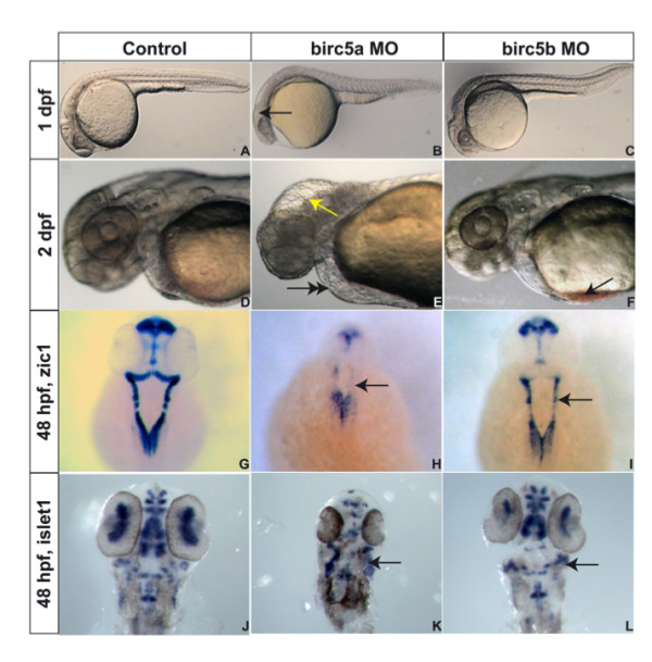
Figure 2.

Birc5 in neurodevelopment. Brightfield microscopy of AB zebrafish embryos. A-F: Embryos oriented with head to left. Lateral views at 1 dpf (A, B, C) and 2 dpf (D, E, F), the latter being higher power views of head region. Depletion of Birc5a results in lack of brain development, revealed at 1 dpf (B, arrow) and 2 dpf (E, arrow), with fluid in 4th ventricle, compared to controls (A, D). At 2 dpf, Birc5a knockdown causes cardiogenic defects and pericardial edema (E, double arrow), not observed in controls (D). Majority of Birc5b depleted embryos do not exhibit phenotypic abnormalities under brightfield microscopy at 1 dpf (C) compared to controls (A). At 2 dpf, Birc5b knockdown embryos have smaller head and brain, and accumulate blood in the sinus venosus (F, arrow). Zic1 expression to detect neural tissue, is decreased by Birc5a depletion (H, arrow), compared to controls (G). A similar but less dramatic diminution of Zic1 expression is observed with morpholino knockdown of Birc5b (I, arrow). Depletion of Birc5a also induces disorganization of motor neurons, detected by expression of islet1 (K, arrow), compared to controls (J). Birc5b morphants exhibit less severe but still evident, suppression of islet1 expression (L, arrow).
