
Figure 1. Panx-1 hemichannels are not activated by cell swelling.
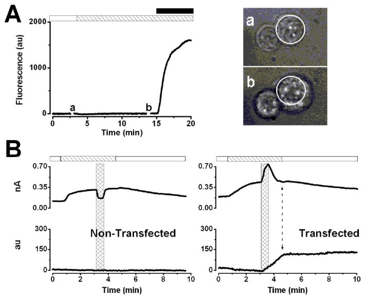
A.- Time course of fluorescence intensity of a representative P2X7-transfected HEK-293 cell (n=5) exposed to Low Tonicity TEACl 190 solution containing EtBr (striped bar). Stimulation with 0.5 mM ATP (black bar) induced EtBr-uptake (end of the trace). Cell images (right) were taken at the time indicated by a and b in the time course. Dotted circles indicate the cell perimeter before the hypotonic challenge. B.- Simultaneous recording of IClswell at +80 mV (upper traces) and fluorescence intensity (lower traces) in non-transfected (n=4) and P2X7-transfected cells (n=4). Cells were exposed to hypotonic TEACl solution containing EtBr (striped bar) and then to 5 mM ATP (hash bars) in a hypotonic solution during 30 s. ATP was washed out using a hypotonic solution, and then ICl,swell was turned off by applying a hypertonic solution (white bar). The double head arrow in the lower time course of the right panel indicates the end of the fluorescence increase.