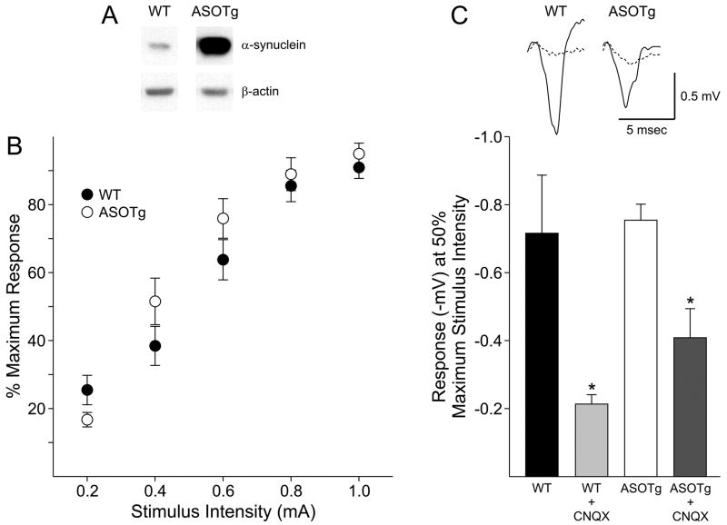
Figure 1. Basal synaptic transmission is unchanged in corticostriatal slices from ASOTg mice and is mainly glutamatergic.
A. Western immunoblotting confirmed elevated expression of human α-synuclein in ASOTg forebrain. Density values (mean ± SEM) for α-synuclein were normalized to β-actin in the same lane (N = 4 each, Student t-test, P < 0.05). B. To generate input-output relationships, the amplitudes of field potentials (-mV) were measured in response to a series of increasing intensities in slices. To standardize responses, the % maximum response (where 100% occurred at the highest stimulus intensity) was plotted against stimulus intensity. No significant differences were evident between WT and ASOTg responses (P = 0.588). C. Basal synaptic transmission recorded at 50% maximum stimulus intensity was significantly reduced in both WT and ASOTg slices treated with CNQX (10 μM, 5 min) to inhibit AMPA/kainate glutamate receptors (* P = 0.02). There was no difference in % change in responses for CNQX-treated WT relative to CNQX-treated ASOTg slices (P = 0.41). Responses shown are averaged synaptic field potentials recorded in WT and ASOTg slices either untreated or treated with CNQX (dashed line).

