
Fig. 12.
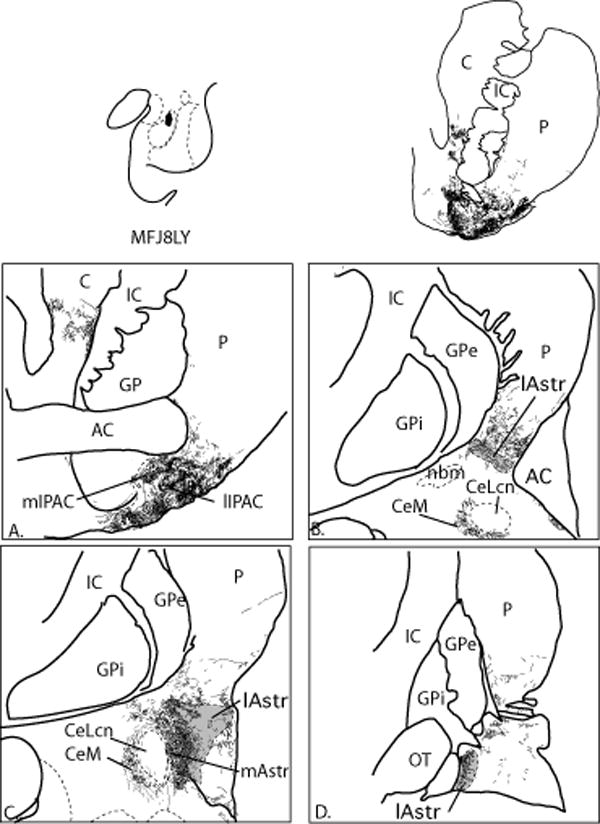
A-D. Distribution of anterogradely labeled fibers resulting from an injection confined to the ABmc (case J8LY). Labeled fibers densely innervate the CeM and all four transition zones, but avoid the CeLcn. The distribution of labeled fibers is in the ventral and lateral shell of the ventral striatum is shown for comparison. Labeled fibers also extend into the ventromedial caudate and putamen. Gray zones depict the CaBP-poor lateral amygdalostriatal area.