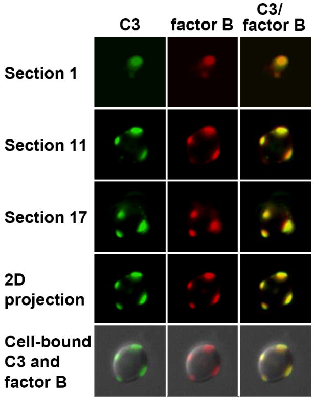
FIG. 5.

Antimannan Fab M1-mediated formation of the alternative pathway C3 convertase on the cell surface of C. albicans. Yeast cells were incubated for 3 min in EGTA-treated yeast-absorbed normal human serum supplemented with 10 μg antimannan Fab M1 per ml and washed. Cell-bound C3 molecules were detected by green FITC-labeled goat anti-human C3 antibody (left, green) and cell-bound factor B molecules were detected by mouse anti-human factor B antibody and red Alexa 594-labeled goat anti-mouse antibody (middle, red). The green and red images were merged to visualize the distribution of bound C3 and factor B on the cell surface (right, yellow). A stack of 20 green or red images were acquired through the Z-axis of a yeast cell and digitally deconvolved. Binding patterns for C3, factor B, or C3-factor B are shown in three individual optical sections (1st, 11th, and 17th) that span the Z-axis of the cell or as a projected 2D image of the 20 sections either by itself or on a DIC image of the cell.
