
Fig. 1.
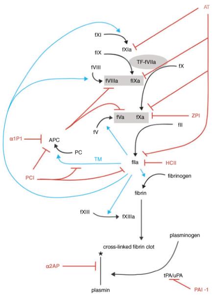
Serpin regulation of coagulation, protein C and fibrinolytic pathways. Serpins and inhibitory functions are shown in red, thrombin activity is shown in cyan. Prothrombinase and tenase complexes are shown in gray boxes. Coagulation is initiated by the exposure of tissue factor to factor VIIa shown in a gray oval. The symbol * indicates degradation. Necessary cofactors, Ca++, phospholipids, proteins S and Z, vitronectin and GAGs are not shown to maintain the simplicity of the schematic.