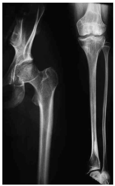
Figure 2.
Radiographs of a case diagnosed as a possible ‘MED variant’ by the expert panel (ESDN-00049, 16 years). The small size and irregular shape of the femoral head, and the flattened appearance of the femoral and tibial condyles are consistent with the post-pubertal changes seen in MED. The slender appearance of the long bones was not considered typical of MED. An Asp385Asn mutation in COMP was identified in this patient.

