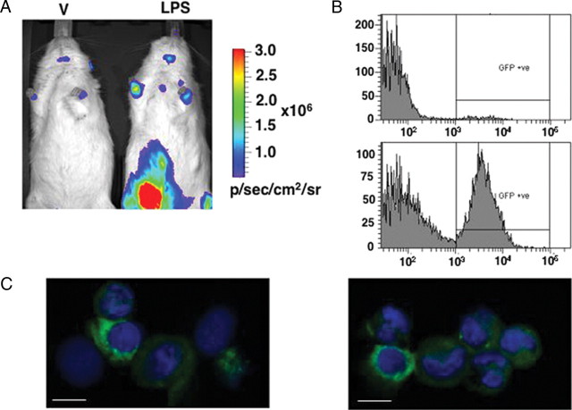
Fig. 6.
In vivo and ex vivo imaging of reporter genes in LPS-treated PRL-Luc/d2eGFP double-transgenic rats. A, In vivo imaging of vehicle (V) and LPS-treated rats. The rats were injected i.p either with physiological saline or with 3 mg/ml LPS and imaged 16 h after the treatment. The images were acquired with an IVIS Spectrum (Caliper Life Sciences): 30 sec integration time, Bin (HR) 8, FOV 19.6, f1. B, Flow cytometric analysis of d2eGFP-positive cells from the peritoneal exudate of a vehicle and a LPS-treated double-transgenic rat. C, Confocal imaging of cytocentrifuge preparation of cells from the peritoneal exudate of LPS-treated double-transgenic rats. Scale bar, 10 μm.

