
Figure 2. Increase in MAGE-11 protein during CWR22 prostate cancer progression.
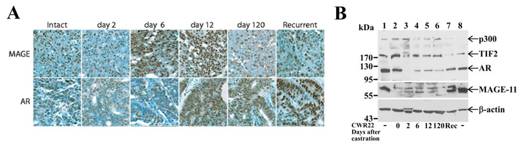
(A) MAGE-11 and AR immunostaining was analyzed in formalin-fixed, paraffin-embedded sections of the CWR22 xenograft excised from intact non-castrated mice, and on days 2, 6, 12 and 120 after castration and longer for the castration-recurrent tumor, using MAGE-11 antibody MagAb94-108 (8 µg/ml) and AR PG21 (Upstate, 1:150 dilution). Brown reaction product indicates immunoreactivity against a toluidine blue counterstain. Original magnification 400X. (B) Immunoblots of MAGE-11, AR, TIF2 and p300 were assayed using CWR22 xenograft extracts prepared from intact-7 (day 0, lane 2), day 2 castrate-3 (lane 3), day 6 castrate-2 (lane 4), day 12 castrate-2 (lane 5), day 120 castrate-3 (lane 6) and castration-recurrent-4 (Rec), according to the numbering of RNA analysis in Fig. 1. Protein extracts (100 µg/lane) were analyzed using antibodies as described in Methods. Combined extracts from COS cells transfected with pSG5-MAGE-11, pCMV-AR, pSG5-TIF and pSG5-HA-p300 served as protein controls (lanes 1 and 8) and endogenous β-actin served as a loading control.