
Figure 3. MAGE-11 and AR expression in clinical specimens of benign and malignant prostate.
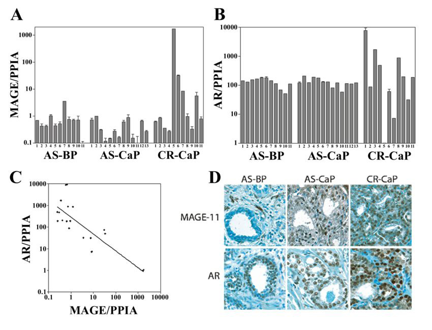
(A) MAGE- 11 and (B) AR mRNA levels relative to peptidylprolyl isomerase A (PPIA) are shown on a semi-log plot determined using quantitative RT-PCR from clinical specimens of androgen-stimulated benign prostate (AS-BP), androgen-stimulated prostate cancer (AS-CaP), and castration-recurrent prostate cancer (CR-CaP). (C) Inverse relationship between AR and MAGE-11 mRNA levels in patient specimens of castration-recurrent prostate cancer. AR and MAGE-11 mRNA levels were determined in duplicate using quantitative PCR of RNA extracted from castration-recurrent prostate cancer specimens. Each of the duplicate data points from the analysis of castration-recurrent specimens in Fig. 2A and B were used to construct the graph. Correlation coefficient R2 = 0.66. (D) MAGE-11 and AR immunostaining of benign and malignant human prostate tissue was performed using formalin-fixed, paraffin-embedded sections of androgen-stimulated benign prostate (AS-BP), androgen-stimulated prostate cancer (AS-CaP), and castration-recurrent prostate cancer (CR-CaP), using MAGE-11 antibody MagAb94-108 (8 µg/ml) and AR antibody PG21 (1:150 dilution). Original magnification 400X.