
Figure 6. MAGE-11 expression in benign and malignant cell lines.
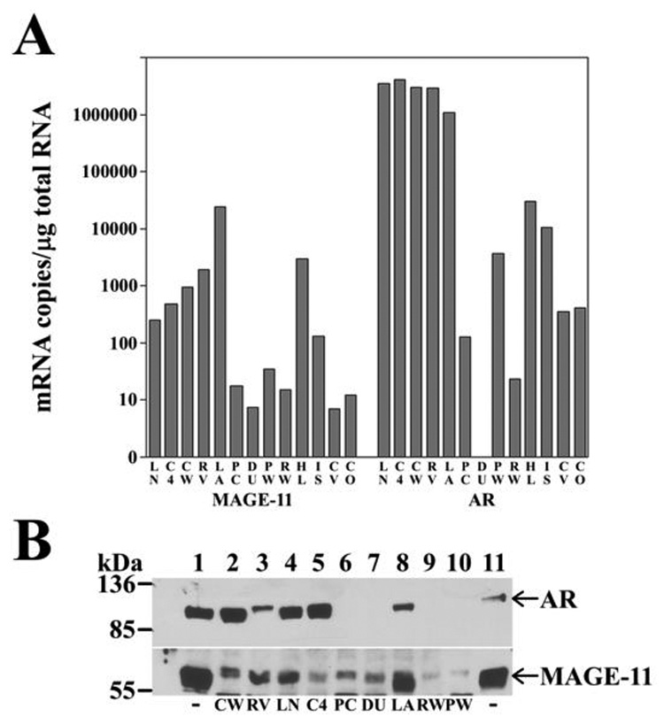
(A) MAGE-11 and AR mRNA levels were determined using quantitative PCR for human prostate cancer cell lines LNCaP (LN), LNCaP-C4-2 (C4), CWRR1 (CW), CWR22-RV1 (RV), LAPC-4 (LA), PC-3 (PC), DU145 (DU), benign human prostate cell lines PWR- 1E (PW) and RWPE-2 (RW), human cervical carcinoma HeLa (HL), human endometrial cancer Ishikawa (IS), monkey kidney CV1 (CV) and COS-1 cells (CO). mRNA amounts were normalized to 1 µg total RNA. (B) Immunoblot of MAGE-11 and AR is shown for extracts (50 µg protein/lane) of CWR-R1 (lane 2), CWR22-RV1 (lane 3), LNCaP (lane 4), LNCaP-C4-2 (lane 5), PC-3 (lane 6), DU145 (lane 7), LAPC-4 (lane 8), RWPE-2 (lane 9) and PWR-1E (lane 10), with abbreviations as in (A). Cells were cultured in serum containing media and analyzed on a 10% acrylamide gel containing SDS. The blot was probed using AR32 (2 µg/lane, top panel) and combined MAGE-11 antibodies 94–108, 59–79 and 13–26 (10 µg/ml each, bottom panel). Combined extracts of COS cells transfected with pSG5-MAGE-11 and pCMV-AR (lanes 1 and 11) were included as controls.