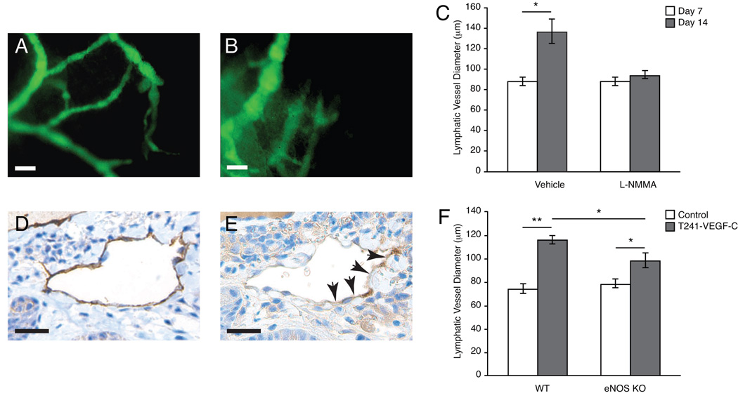
Figure 3. Effect of pharmacologic and genetic NOS-inhibition on peritumor lymphatic hyperplasia.
(A-C) Representative fluorescence images after FITC-Dextran lymphangiography. Animals received PBS or L-NMMA treatment starting on 7 days after T241-VEGF-C tumor implantation. Fourteen days after tumor implantation, lymphangiography was repeated and revealed that hyperplasic peritumor lymphatics in control animals (A) and attenuation in peritumor lymphatics hyperplasia in L-NMMA treated animals (B). (C) Quantification of the lymphatic vessel diameters after lymphangiography showed significantly reduced hyperplasia after 7 days of L-NMMA treatment. (D, E) Immunohistochemistry for LYVE-1 (D) and eNOS (E) shows that eNOS is expressed in peritumor lymphatics. Scale bars represent 100µm. (F) Rhodamine-Dextran lymphangiography revealed a significant reduction of peritumoral lymphatic hyperplasia in eNOS−/− mice implanted with T241-VEGF-C-GFP tumors compared to tumors implanted in wt mice. Normal ear lymphatics (control) had similar diameters. *P<0.05, **P<0.01, Student t test.

