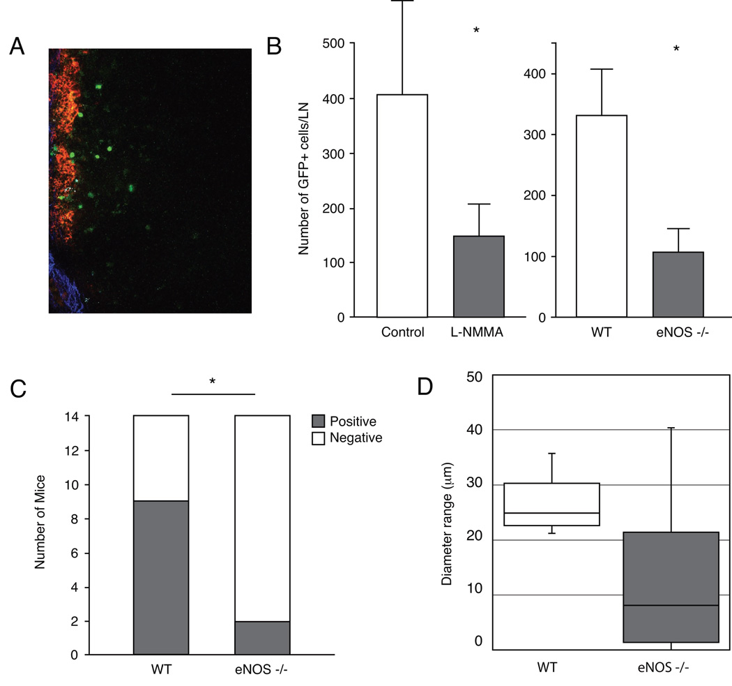
Figure 4. Effect of pharmacologic and genetic eNOS-inhibition on lymph node metastasis.
(A) Image of a lymph node where seeded GFP+ T241-VEGF-C tumor cells can be seen in green. Red color indicates the Rhodamine-Dextran lymphangiography and the blue color indicates the collagen capsule of the lymphnode as visualized by second harmonic generation. (B) Animals received L-NMMA treatment starting 7 days after T241-VEGF-C-GFP ear tumor implantation. Fourteen days after implantation, GFP+ tumor cells in the cervical lymph node were quantified using multiphoton laser-scanning microscopy (9). L-NMMA significantly inhibited arrival of metastatic GFP+ tumor cells to the draining lymph node. Multiphoton laser-scanning microscopy also revealed a significant reduction of metastatic GFP+ tumor cell arrival to the draining lymph node 7 days after T241-VEGF-C-GFP ear tumor implantation in wt or eNOS−/− mice. (C) Genetic eNOS inhibition attenuates formation of clinical lymphatic metastasis. Fourteen days after B16F10 tumor implantation in mouse ears, cervical lymph nodes were inspected for the presence of metastasis lesions. Number of metastasis was significantly reduced in eNOS−/− mice when compared to wt mice. (D) Although statistical significance was not reached, LYVE 1 immunohistochemistry showed hyperplastic B16F10 peritumor lymphatic vessels in control mice and moderately hyperplastic lymphatic vessels in eNOS−/− mice. *P<0.05, Student t test (A and D), Mann-Whitney-U-test (B), Fisher’s exact test (C).

