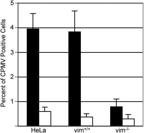
Figure 3. Cell-surface expression of vimentin promotes interaction with CPMV.
Flow cytometry of surface vimentin-expressing cells HeLa and MFT-6 vim+/+, and vimentin negative MFT-16 vim−/− cell types incubated with (black bars) and without (white bars) 105 CPMV particles per cell for 30 minutes at 37°C in respective growth media. Bars represent mean+/−s.d. of triplicate samples.

