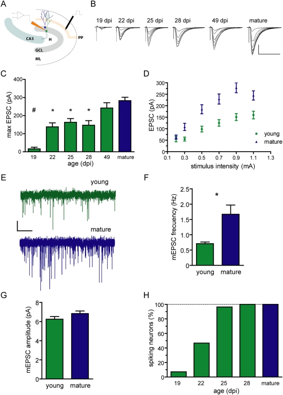
Figure 2. Spikes in young neurons elicited by weak glutamatergic inputs.
(A) Illustration depicting electrophysiological recordings of postsynaptic responses in acute hippocampal slices. Stimulation was performed in the medial perforant path (pp), and whole-cell recordings were obtained in young (green) or mature (blue) granule cells. GCL, granule cell layer; ML, molecular layer; H, hilus. (B) Example traces of EPSCs evoked by increasing stimulus strength (0.2–1 mA, 50 µs) obtained from DGCs of different developmental stages (shown on the top). Each trace is an average of five epochs. Scale bars: 100 pA, 50 ms. (C) Maximal peak EPSC amplitude for young and mature DGCs. (#) denotes p<0.001, and (*) denotes p<0.05 when compared to mature, as analyzed by ANOVA followed by a Bonferroni's post-hoc comparison, with N = 9 (19 dpi), 9 (22 dpi), 14 (25 dpi), 10 (28 dpi), 10 (49 dpi) and 41 (mature). (D) Peak EPSC amplitude vs. stimulus intensity in young (21–29 dpi) and mature DGCs. Two-way ANOVA revealed a significant effect of both neuronal age and stimulus intensity (p<0.0001 for both), with N = 27 for both groups. (E) Representative traces of miniature postsynaptic currents obtained in presence of 0.5 µM tetrodotoxin. Scale bars: 2.5 pA, 5 s. (F, G) Frequency and amplitude of mEPSCs. (*) denotes p = 0.016; for amplitude p = 0.14; N = 8 for both. (H) Fraction of spiking neurons as a function of age. Spiking was measured in whole-cell current clamp and was elicited by stimuli that rendered maximal EPSC amplitudes, with N = 14 (19 dpi), 15 (22 dpi), 28 (25 dpi), 21 (28 dpi) and 59 (mature).

