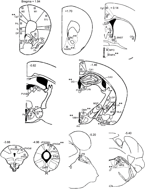
Figure 1. Schematic diagram showing the 59 areas in which c-Fos expression was quantified.
Levels are based on the atlas of Franklin and Paxinos (1997). Squares indicate the placement of grids for counting of c-Fos positive cells. Asterisks indicate the regions in which HAB mice showed changes in OA-induced c-Fos expression as compared to LABs. AcB, nucleus (n.) accumbens; AcBc, n. accumbens core; AcBsh, n. accumbens shell; ACo, anterior cortical n. of the amygdala; AD, anterodorsal thalamic n.; AH, anterior hypothalamic area; Arc, arcuate hypothalamic nucleus; BlA, basolateral n. of the amygdala; BNST, bed n. of the stria terminalis; CA1, CA1 field of the hippocampus; CA3, CA3 field of the hippocampus; CeA, central n. of the amygdala; Cg 1, cingulate ctx (area1); Cg 2, cingulate ctx (area2); Cl, Claustrum; CPu, caudate putamen; cPAGdl, caudal dorsolateral periaqueductal gray; cPAGdm, caudal dorsomedial periaqueductal gray; cPAGl, caudal lateral periaqueductal gray; cPAGvl, caudal ventrolateral periaqueductal gray; DEn, Endopiriform ctx, dorsal; DG, dentate gyrus; DMH, dorsomedial hypothalamic n.; DP, dorsal peduncular nucleus; DR, dorsal raphe n.; GI, granular insular ctx; IL, infralimbic ctx; LA, lateral n. of the amygdala; LC, locus coeruleus; LGP, lateral globus pallidus; LH, lateral hypothalamic area; LHb, lateral habenular n.; LPB, lateral parabrachial n.; LSD, lateral septal n. (dorsal); LSI, lateral septal n. (intermediate); LSV, lateral septal n. (ventral); M1, primary motor ctx; M2, secondary motor ctx; MeA, medial amygdala; MGP, medial globus pallidus; MO, medial orbital cortex; MPA, medial preoptic area; MPO, medial preoptic n.; MPB, medial parabrachial n.; PE, periventricular n; Pir, piriform ctx; PLCo, posterolateral cortical n. of the amygdala; PrL, prelimbic ctx; PV, paraventricular thalamic n.; PVA, paraventricular thalamic n. (anterior); PVN, paraventricular hypothalamic n.; rPAGdl, rostral dorsolateral periaqueductal gray; rPAGdm, rostral dorsomedial periaqueductal gray; rPAGl, rostral lateral periaqueductal gray; RSA, retrosplenial agranular ctx; RSG, retrosplenial granular ctx; S1J, primary somatosensory cortex, jaw region; VMH, ventromedial hypothalamic n.

