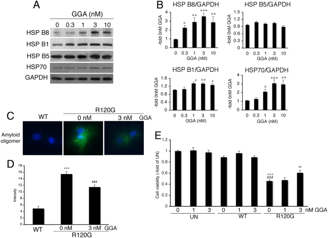
Figure 1. Induction of small HSPs by GGA in neonatal rat cardiomyocytes.
(A) Western blot analysis. GGA induced the expression levels of small HSPs in a dose-dependent manner. (B) Quantitative analysis of small HSP expression (n = 4). Values are the -fold increase relative to cardiomyocyte cultures treated with vehicle (0 nM of GGA) whose values are arbitrarily set to 1. * p<0.05, ** p<0.01 and *** p<0.001 vs. 0 nM of GGA. (C) Representative pictures of immunohistochemical analyses in the HSPB5 R120G-infected cardiomyocytes. Cardiomyocytes were infected with the adenovirus vector containing the wild-type HSPB5 (WT) or mutant HSPB5 R120G (R120G). An amyloid oligomer (green) was detected by the anti-oligomer antibody as described in Materials and Methods. Nuclei were stained with DAPI. (D) Quantitative analysis of the amyloid oligomer. Amyloid oligomer levels were measured by fluorescence intensity (n = 4). *** p<0.001 vs. cardiomyocytes infected with WT, and ### p<0.001 vs. cardiomyocytes infected with R120G. (E) Protective effects of GGA on the cellular viability of the HSPB5 R120G-infected cardiomyocytes. Cellular viability was determined by the MTT assay. Values are the -fold increase relative to untreated cardiomyocyte cultures whose values are arbitrarily set to 1 (n = 6). *** p<0.001 vs. untreated cardiomyocytes (UN), ### p<0.001 vs. cardiomyocytes infected with WT, and aa p<0.01 vs. cardiomyocytes infected with R120G.

