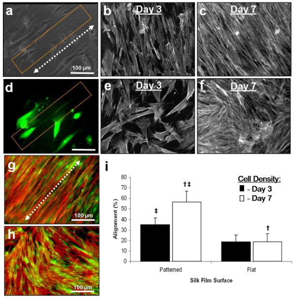
Fig. 4.
(a) Phase contrast image of a GFP-rCF cellular process highly aligned along the patterned silk film groove axis (arrow) after 4 days in culture. (b) Fluorescent image of the same cellular process indicating true cellular component. Actin staining for both (g) patterned and (h) flat silk film surfaces after 10 days in culture. Images are in false color; red indicates stained actin filaments and green represents GFP fluorescence. (i) A statistical significance between actin alignment on patterned and flat silk film surfaces was found († indicates statistical difference; p<0.05; error bars=SD; n=3). Fluorescent images of actin filament organization for different culture time points of GFP-rCFs seeded upon (b)(c) patterned and (e)(f) flat silk film surfaces. (b)(e) Lower cell densities exhibited greater cell spreading. At higher cell densities the cells became more compact and exhibited greater aligned organization along the silk film groove axis (c) as compared to flat silk film surfaces (f). (i) Cell density affects actin filament organization on patterned silk film surfaces. (i) A statistically significant increase in actin filament alignment is observed at increased cell densities († indicates statistical difference; p<0.05; error bars = SD; n=3). No affect on actin filament alignment was observed between cell density groups on flat silk film surfaces.

