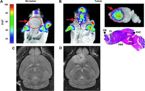
Figure 4.
BLI and MRI are useful tools for visualization of tumors in vivo. (A) BLI image of non-tumor-bearing Gli-Luc mouse (red arrow and circle points to the brain, where there is no signal). (B) In vivo BLI image of PDGF-induced glioma injected in the SVZ, with ex vivo sagittal image of the same tumor with corresponding H&E. FL indicates frontal lobe; OB, olfactory bulb. (C and D) T2-weighted images of non-tumor- and tumor-bearing mice (injected with PDGF in the SVZ), respectively.

