Figure 1.
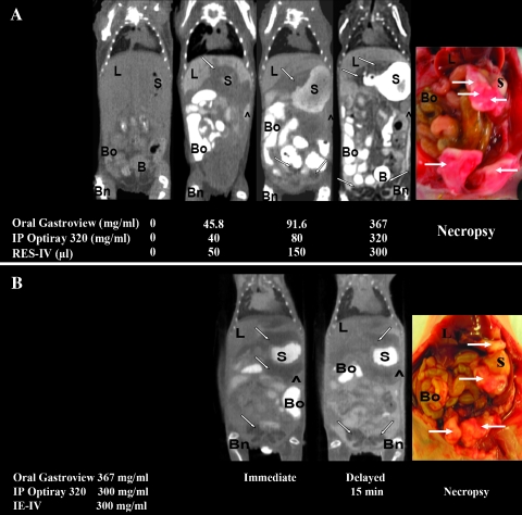
(A) Representative white light and in vivo CT images of IP Hey A8 tumors before and after administration of different doses of oral, IP, and RES-IV contrasts. (B) Representative immediate and delayed (15 minutes) in vivo CT images and white light images of IP Hey A8 tumors in mice administered oral, IP, and IE-IV contrasts. In the pre-mice, without any contrast, tumor and other organs are not well visualized. Upon administering contrasts, tumor and organs are better visualized. Tumor appears as areas of low attenuation compared with adjacent structures. ^ indicates peritoneum; B, bladder; Bn, bone; Bo, bowel; L, liver; S, stomach.

