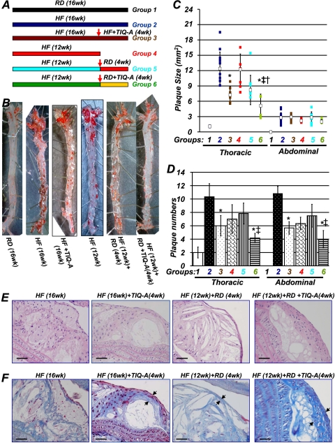
Fig. 1.
PARP inhibition by TIQ-A promotes atherosclerotic plaque regression in an animal model of atherosclerosis. A, diagram of the study. ApoE(-/-) mice (n = 60) were fed an HF diet for 12 weeks, after which one group was sacrificed immediately (n = 12), and the remaining were divided into four groups of at least 12 mice each. One of these groups was continued with the high-fat diet, one group was continued with the high-fat diet and received 3 mg/kg TIQ-A (3 times/week), and the other two were switched to an RD for 4 additional weeks, receiving 3 mg/kg TIQ-A (3 times/week) [HF (12 weeks) + RD + TIQ-A (4 weeks)] (n = 12) or vehicle [HF (12 weeks) + RD (4 weeks)] (n = 12); the control group received a regular diet throughout the 16 weeks (RD; 16 weeks). B, aortas from the different experimental groups were formalin (phosphate-buffered saline)-perfused, and atherosclerotic plaques were visualized by en face Oil Red O staining. Plaque size (C) and number (D) were determined as described previously (Oumouna-Benachour et al., 2007a). *, difference from ApoE(-/-) mice fed a high-fat diet for 16 weeks; ‡, difference from ApoE(-/-) mice fed a high-fat diet for 12 weeks, followed by 4 weeks of the regular diet; p < 0.01; open squares, medians. †, difference from ApoE(-/-) mice fed a high-fat diet and administered TIQ-A for 4 weeks. E, hematoxylin and eosin staining of representative plaques from the different experimental groups; bar, 5 μm. F, trichrome staining of representative plaques from the different experimental groups; bar, 5 μm. Arrows, TIQ-A not only increased collagen content but also caused thickening of fibrous caps.

