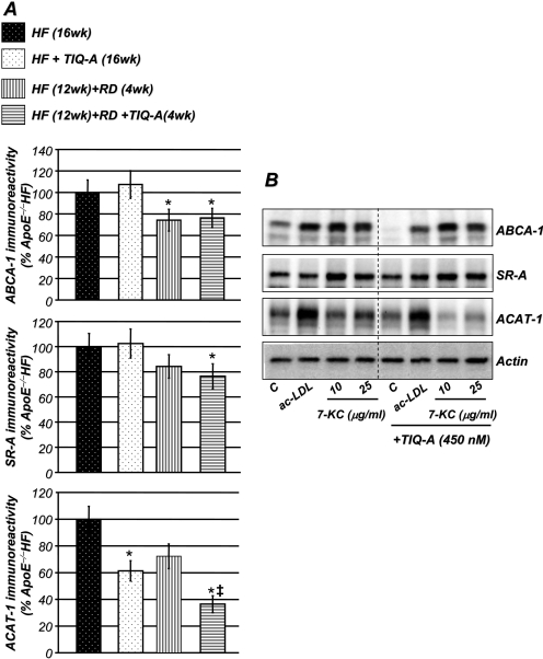
Fig. 6.
Effects of TIQ-A on ABCA-1, SR-A, and ACAT-1 expression in vivo and in vitro. A, immunohistochemistry analysis with antibodies to ABCA-1, SR-A, or ACAT-1 of plaques from the different experimental groups; quantitation of immunoreactivity was conducted using Image-Pro Plus software and expressed as percentage immunoreactivity detected in plaques of ApoE(-/-) fed high-fat diet for 16 weeks. *, difference from ApoE(-/-) mice fed the high-fat diet for 16 weeks; ‡, difference from ApoE(-/-) mice fed the high-fat diet for 12 weeks followed by 4 weeks of regular diet; P < 0.01. B, foam cells were generated through an incubation with acetylated-LDL (10 μg/ml) as described previously (Oumouna-Benachour et al., 2007a). Cells were then treated with acetylated-LDL (10 μg/ml) or 7-ketocholesterol for an additional 12 h. Cell extracts were prepared and subjected to immunoblot analysis with antibodies to ABCA-1, SR-A, ACAT-1, or actin. These are representative blots for four different experiments.

