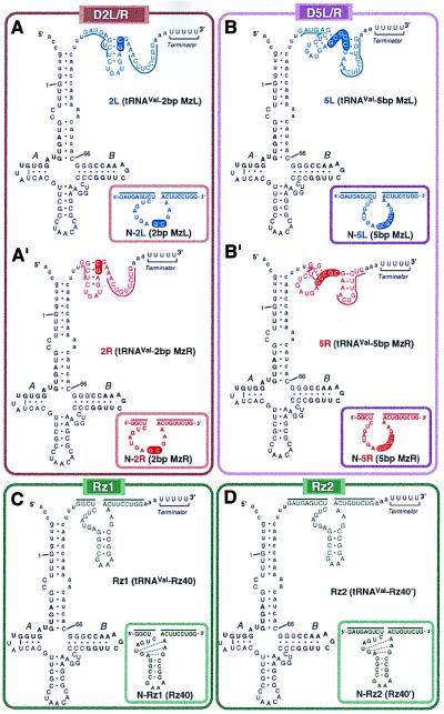
Figure 2.
Predicted secondary structures, based on calculations by the mulfold program (Biocomputing Office, Biology Department, Indiana University, Bloomington, IN), of tRNAVal-enzymes. The human tRNAVal sequence, including the binding sites of transcription factor TFIIIC (labeled A and B, corresponding to the A and B box regions), is indicated in uppercase letters with numbering from 1 to 66. Extra sequences that were inserted artificially are indicated by lowercase letters. The sequences of L (maxizyme left) and R (maxizyme right) are shown in blue and red, respectively, and the sequences of standard ribozymes (C and D) are shown in green. Thick lines indicate the substrate-binding regions of the enzymes. The common stem II regions of the dimeric maxizymes (in A and B) are indicated by outlined letters within a solid ellipse.

