
Figure 3. Low numbers of peripheral T cells in CD4Cre/R-DTA mice.
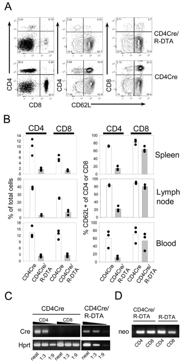
A) Mesenteric lymph node cells from CD4Cre/R-DTA and CD4Cre control mice were stained with anti-CD4, anti-CD8 and anti-CD62L antibodies and analyzed by flow cytometry. B) Blood, spleen and mesenteric lymph nodes of three individual mice were analyzed for the frequency of CD4+ and CD8+ T cells among total cells (left graphs) and the frequency of CD62L+ cells among CD4+ and CD8+ T cells (right graphs). Results are representative of several independent experiments. C) Semiquantitative RT-PCR analysis for Cre- and Hprt-expression of sorted CD4+ and CD8+ T cells from CD4Cre mice (left) and sorted CD4+ T cells from CD4Cre/R-DTA mice (right). D) Genomic PCR of sorted CD4+ and CD8+ T cells from CD4Cre and CD4Cre/R-DTA mice to determine the presence or absence of the loxP flanked neomycin resistance gene.