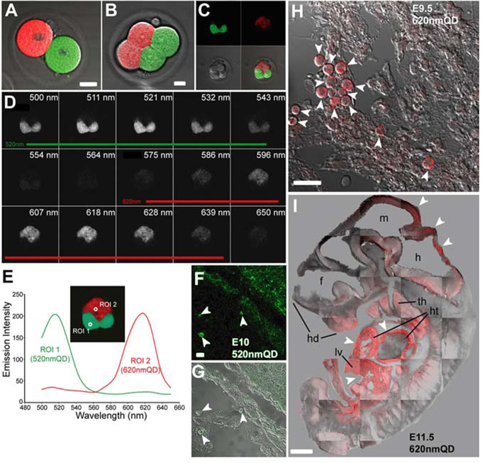
Fig. 2. Quantum dot (QD) labeling of early mouse embryos does not alter fetal development.
A: Two-cell stage mouse embryos injected with 520-nm and 620-nm QDs in vitro were allowed to divide for 1–2 days in culture. B,C: Four-cell (B) and eight-cell (C) stage embryos developed with compartmentalized segregation of QD into daughter cells during development demonstrating there was no mixing of QD during cell division. D: Lambda stack image illustrates the spectral characteristics of each QD. E: Emission fingerprints of 520-nm and 620-nm QD. F–I: One- and two-cell stage 520-nm or 620-nm QD-injected mouse embryos were implanted into pseudo-pregnant dams for 9.5 to 11.5 days of in utero development. All embryonic tissues appeared to contain QDs, although there was uneven labeling with some tissues containing a higher density of QD. E–G: For example, cells in the umbilical region of an embryonic day (E) 10 fetus injected with 520-nm QD (arrowheads in F,G) had more QD labeling than the underlying epithelial cells. H: Another E9.5 fetus labeled at the one-cell stage with 620-nm QD, exhibited some heavily labeled ventricular pericardial cells (arrowheads) while other neighboring cells possessed much lower QD labeling. I: In an E11.5 embryo where 620-nm QD were loaded into one-cell at the two-cell stage, there was asymmetrical labeling of body tissues by QDs. There was much higher labeling (arrowheads) in the liver (lv), cardiac primordium (ht), thymus (th), and hindbrain (h) compared with other tissues. hd, head; f; forebrain; m, midbrain. Scale bars = 5 µm in A,B; 15 µm in F,G; 20 µ m in H; 0.5 mm in I.

