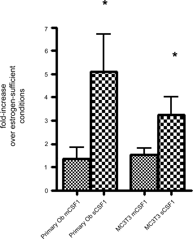
Fig. 4.
Effect of estrogen withdrawal for 24 h on levels of mCSF1 and sCSF1 transcript expression in primary murine osteoblasts isolated from the calvariae of female pups (left two bars) and in MC3T3E1 cells (right two bars). Levels of CSF-1 isoform expression were quantified by quantitative RT-PCR and expressed as fold change compared with estrogen-replete parallel cultures. Data reflect mean results from 4–7 experiments. *P < 0.05, compared with estrogen-replete culture conditions. +P < 0.05, compared to wild-type controls.

