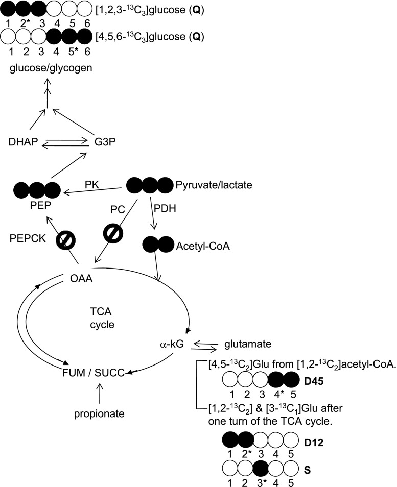
Fig. 4.
Schematic diagram of metabolic pathways and 13C labeling patterns related to reverse flux through pyruvate kinase in skeletal muscle after [U-13C3]lactate administration. The direct conversion of [U-13C3]lactate to [U-13C3]pyruvate followed by reverse flux through pyruvate kinase results exclusively in [1,2,3-13C3]- and [4,5,6-13C3]glucose units in muscle glycogen without other labeling patterns. If phosphoenolpyruvate (PEP) formation from pyruvate occurs through a bypass (pyruvate → oxaloacetate → PEP) as seen in liver (Fig. 6, A and B), various glucose isotopomers including both [1,2,3-13C3]- and [4,5,6-13C3]glucose plus other distinctive labeling patterns such as [1,2-13C2]glucose, etc. would be observed due to rearrangements of 13C in the TCA cycle and pyruvate cycling. The glutamate analysis of the tissue showed only [4,5-13C2]-, [1,2-13C2]-, and excess [3-13C1]glutamate isotopomers. This informs that [U-13C3]pyruvate incorporated into the TCA cycle only through pyruvate dehydrogenase but not through the pyruvate carboxylase pathway. PDH, pyruvate dehydrogenase; PK, pyruvate kinase; PC, pyruvate carboxylase; PEPCK, phosphoenolpyruvate carboxykinase; OAA, oxaloacetate; G3P, glyceraldehyde 3-phosphate; DHAP, dihydroxyacetone phosphate; a-kG, alpha-ketoglutarate. Open circle, 12C; filled circle, 13C.

