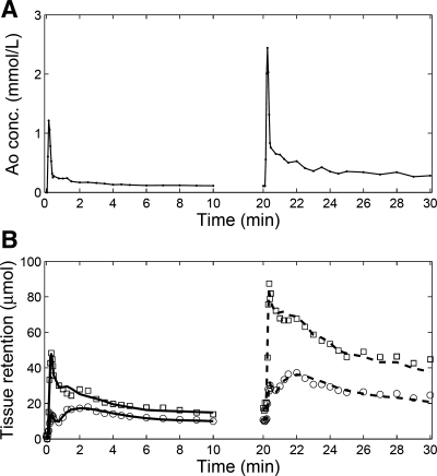
Fig. 4.
Measured double-injection MRR data and model fits for a representative kidney. A: tracer concentrations measured from an aortic region of interest. B: tracer retention in cortex is represented by squares and that from medulla by circles. The first-injection data (0–10 min) were fitted using the model described by equations 3-5 (solid lines), with results GFR1 = 43.8 ml/min, RPF1 = 208.4 ml/min. The second-injection data (20–30 min) were fitted using the model described in equations 3 and 6 (dashed lines), with results GFR2 = 44.9 ml/min, RPF2 = 177.6 ml/min. In this case, GFR2 − GFR1 = 1.1 ml/min, while RPF2 − RPF1 = −30.8 ml/min. In the absence of renovascular disease, the expected GFR2 − GFR1 value is 0.

